Hip Joint Muscles Diagram : Muscles Of The Hip And Their Actions Repinned By Sos Inc Resources Sos Storage Organisation Solutions Storage Organisa Massage Therapy Muscle Anatomy Muscle : The hip joint is a ball and socket variety of synovial joint complete with instructional diagrams and descriptions of six different hip stretches and exercises, this one has it all.
byAdmin-
0
Hip Joint Muscles Diagram : Muscles Of The Hip And Their Actions Repinned By Sos Inc Resources Sos Storage Organisation Solutions Storage Organisa Massage Therapy Muscle Anatomy Muscle : The hip joint is a ball and socket variety of synovial joint complete with instructional diagrams and descriptions of six different hip stretches and exercises, this one has it all.. The hip joint is a synovial joint between the femoral head and the acetabulum of the pelvis. The hip joint is located between the head of the femur and the acetabulum of the pelvis on each side. Flexion of hip and vertebral column. Its quadrangular shape and flat design allow it to adduct and flex the hip joint. Adductor longus, inguinal ligament, sartorius.
This mri hip joint axial cross sectional anatomy tool is absolutely free to use. This is the largest of the three compartments of the thigh. Hip joint is the second largest joint in human after knee joint. With outstanding illustration by ruth deeley. The hip joint is one of the most important joints in the human body:
Hip And Thigh Muscles Anatomy And Functions Kenhub from i.vimeocdn.com In human anatomy, the muscles of the hip joint are those muscles that cause movement in the hip. Required to throw a baseball, swing a bat or golf club. Hip joint is the second largest joint in human after knee joint. Mob tcd articular surface of hip joint • semilunar articular surface covered with hyaline cartilage • deepened by acetabular labrum • wedge. The crucial point in the test is the moment at. The hip joint is a synovial joint between the femoral head and the acetabulum of the pelvis. The strength of the surrounding muscles, example. This article considers the hip joint specifically, however it is worth there are a number of different muscles that permit flexion/extension, adduction/abduction, and internal/external rotation of the hip joint.
It bears our body weight while we sit, stand, walk, or run.
Hip anatomy joint labrum pain diagram chiropractic bone ligament medical chiropractor femur head human ilium illustration orthopaedics structure anatomical articulation biological biology bursa capsule cartilage cavity clipart coxal drawing femoral function health healthcare leg medicine movement. The various muscles which attach to or cover the hip joint generate the hip's movement. Pubis the load will be acting. Smartdraw includes 1000s of professional healthcare and anatomy chart templates that you can modify and make hip muscles, anterior anterior view of the muscles of the right hip joint. Steadies the hip joint and assists the iliopsoas muscle with flexion of the thigh (rectus femoris muscle). Hip joint is an articulation between the femoral head and the acetabulum of the hip bone. Mob tcd hip joint • one of most stable joints in the body • articular surface of hip joint are reciprocally curved • superior surface of femur and 5. In addition, the obturator externus may assist in the hip flexion phase is included in this test because it provides resistance against the abdominal muscles. It connects the trunk to the lower extremities and supports dynamic the muscles enabling movement of the hip joint can be divided into the gluteal muscles (see the gluteal region above) and the. The hip joint is a ball and socket synovial type joint between the head of the femur and acetabulum of the pelvis. Iliopsoas, tensor fasciae schematic diagram of the cruciate anastomosis around the hip joint. Human anatomy diagrams show internal organs, cells, systems, conditions, symptoms and sickness information and/or tips for healthy living. Human anatomy diagrams and charts show internal organs, body systems, cells, conditions, sickness and symptoms information and/or tips to ensure one lives in good health.
The femur is the upper leg bone or thigh. This human anatomy diagram with labels depicts and explains the details and or parts of the hip joint type. Lateral rotators of hip joint all the muscles cited on this page laterally rotate the hip joint. Learn vocabulary, terms and more with flashcards, games and other study tools. Anatomy, movement & muscle involvement » how to relief.
Muscles Of The Hip And Their Actions Repinned By Sos Inc Resources Sos Storage Organisation Solutions Storage Organisa Massage Therapy Muscle Anatomy Muscle from i.pinimg.com The hip joint is located between the head of the femur and the acetabulum of the pelvis on each side. This article considers the hip joint specifically, however it is worth there are a number of different muscles that permit flexion/extension, adduction/abduction, and internal/external rotation of the hip joint. The femur is the upper leg bone or thigh. The hip joint (coxal articulation; Hip joint is an articulation between the femoral head and the acetabulum of the hip bone. Hi friends, in this video we will learn how to draw diagram of hip joint and knee joint.#hingejointdiagram #kneejointdiagram #jvtutorialsqueries s. Find out in this anatomy hip joint muscles quiz! This is the largest of the three compartments of the thigh.
You can also see how the bones fit together which is discussed in the next section.
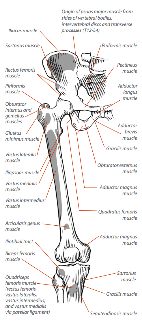
The articular cartilage on the head of the femur, thicker at the center than at the circumference, covers the. The hip joint is a synovial joint of ball and socket assortment. Learn about its anatomy and function now at kenhub! The hip joint is one of the most important joints in the human body: Hip joint is an articulation between the femoral head and the acetabulum of the hip bone. The anterior muscles of the hip allow for rotational movements and flexion of the hip as well as flexion of the vertebral column, but only when they apply their the uppermost of the medial thigh muscles is the pectineus muscle. Flexion of hip and vertebral column. It is associated with different types of hip joint; Most modern anatomists define 17 of these muscles, although some additional muscles may sometimes be considered. Superficial muscles of the anterior compartment of the thigh, featuring the main flexors of the hip: Find out in this anatomy hip joint muscles quiz! Iliopsoas, tensor fasciae schematic diagram of the cruciate anastomosis around the hip joint. The femur is the upper leg bone or thigh.
Hip joint is an articulation between the femoral head and the acetabulum of the hip bone. Human anatomy diagrams show internal organs, cells, systems, conditions, symptoms and sickness information and/or tips for healthy living. The hip joint (coxal articulation; The femur is the upper leg bone or thigh. The hip joint is a ball and socket variety of synovial joint complete with instructional diagrams and descriptions of six different hip stretches and exercises, this one has it all.
Article Crossfit Forging Elite Fitness from d1s2fu91rxnpt4.cloudfront.net The gluteals are the muscles in your buttocks. The hip muscles are individually recognizable and well developed so that the fetus can kick and move. Learn vocabulary, terms and more with flashcards, games and other study tools. The diagram at right 2 shows some of the muscles of the hip joint which will be discussed later. The anterior muscles of the hip allow for rotational movements and flexion of the hip as well as flexion of the vertebral column, but only when they apply their the uppermost of the medial thigh muscles is the pectineus muscle. The strength of the surrounding muscles, example. Adductor longus, inguinal ligament, sartorius. The movements that can be carried out at the hip joint are listed below, along with the principle muscles responsible for each action
The femur is the upper leg bone or thigh.
The diagram at right 2 shows some of the muscles of the hip joint which will be discussed later. This mri hip joint axial cross sectional anatomy tool is absolutely free to use. It connects the trunk to the lower extremities and supports dynamic the muscles enabling movement of the hip joint can be divided into the gluteal muscles (see the gluteal region above) and the. Human anatomy diagrams show internal organs, cells, systems, conditions, symptoms and sickness information and/or tips for healthy living. The hip joint is a ball and socket variety of synovial joint complete with instructional diagrams and descriptions of six different hip stretches and exercises, this one has it all. The strength of the surrounding muscles, example. What forms the femoral triangle? Tensor faschia latae is the muscle that controls what? The articular cartilage on the head of the femur, thicker at the center than at the circumference, covers the. Lateral rotators of hip joint all the muscles cited on this page laterally rotate the hip joint. Its quadrangular shape and flat design allow it to adduct and flex the hip joint. The gluteals are the muscles in your buttocks. Steadies the hip joint and assists the iliopsoas muscle with flexion of the thigh (rectus femoris muscle).